Figure 3.
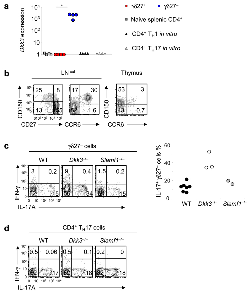
DKK3 and SLAMF1 are molecular determinants of γδ27− T cells. (a) Quantitative RT-PCR analysis of Dkk3 expression in ex vivo CD4+ T cells, γδ27+ T cells and γδ27− T cells (derived from peripheral T cells) and CD4+ Th1 and Th17 cells generated in vitro (all sorted from C57BL/6 mice); results are presented relative to those of Actb (control gene). *P < 0.05 (Mann-Whitney two-tailed test). (b) Surface expression of SLAMF1 (CD150), CD27 and CCR6 on total γδ T cells from cutaneous lymph nodes (LN (cut); left) and thymus (right), assessed by flow cytometry. Numbers in quadrants indicate percent cells in each throughout. (c) Intracellular staining of IFN-γ and IL-17A in γδ27− cells isolated from C57BL/6 wild-type (WT), Dkk3−/− and Slamf1−/− mice (left), and frequency of IL-17+ γδ27− T cells in those mice (right; n = 7 wild-type mice, 3 Dkk3−/− mice and 2 Slamf1−/− mice). (d) Intracellular staining of IFN-γ and IL-17A in Th17 cells differentiated in vitro from CD4+ T cells isolated from WT, Dkk3−/− and Slamf1−/− mice. Each symbol (a,c, right) represents an individual experiment. Data are from one experiment with cells pooled from four mice (a) or one experiment per mouse (c, right) or are representative of three independent experiments with two or three mice in each (b) or two independent experiments with for mice per strain in each (d).

