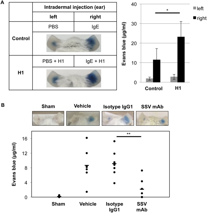
Fig 4. Enhancement and inhibition of IgE-mediated PCA reaction by histone H1 and histone H1-targeted SSV mAb.
(A) PBS and anti-DNP IgE (150 ng/10 μl) with/without calf thymus histone H1 (5 μg) were intradermally injected into the left and right ears, respectively (n = 3 per group). After 24 hrs, DNP-HSA (200 μg) with evans blue solution (1%) was injected intravenously via tail vein to induce anaphylaxis. After 30 minutes, evans blue extravasation in the ears was observed. The pictures are representative of three individuals in each group. Data are representative of three independent experiments and represented as the mean ± S.D. *, P<0.05 versus the control group. (B) PBS and anti-DNP IgE (150 ng/10 μl) were intradermally injected into the left and right ears, respectively. As a sham control (n = 8), PBS was injected intradermally in both left and right ears. After 23 hrs, PBS (vehicle; n = 7), isotype IgG1 (n = 8) or SSV mAb (n = 7) was intravenously injected via tail vein. One hr later, DNP-HSA (200 μg) with evans blue solution (0.5%) was injected intravenously via tail vein to induce anaphylaxis. After 30 minutes, evans blue extravasation in the ears was observed. The pictures are representative of seven to eight individuals in each group. Each symbol indicates an individual mouse, and bars show the mean values. **, P<0.01 versus the isotype IgG1-injected group.

