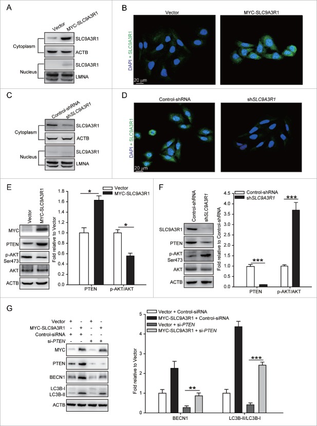
Figure 2.
(See previous page). SLC9A3R1 partially stimulates autophagy through the PTEN-PI3K-AKT1 pathway. (A–D) SLC9A3R1 is mostly distributed in the cytoplasm of breast cancer cells. For panels (A and C), breast cancer cells were harvested, and the subcellular fraction was extracted for immunoblotting. ACTB and LMNA were used as loading controls. For panels (B and D), breast cancer cells in dishes were fixed in 4% paraformaldehyde, stained with fluorochromes, and imaged by confocal microscopy, Scale bar: 20 μm. For panels (A and B), the experiments were carried out in MDA-MB-231 cells stably expressing vector or MYC-SLC9A3R1. For panels (C and D), the experiments were carried out in MCF-7 cells stably expressing control-shRNA or sh-SLC9A3R1. (E) SLC9A3R1 stimulates the PTEN-PI3K-AKT1 pathway in MDA-MB-231 cells. MDA-MB-231 cells stably expressing vector or MYC-SLC9A3R1 were harvested, and the expression of the indicated proteins was determined by immunoblotting. The value for the vector was set to 1.0 and the other values were normalized. (F) Silencing of SLC9A3R1 suppresses the PTEN-PI3K-AKT1 pathway in MCF-7 cells. MCF-7 cells stably expressing control-shRNA or sh-SLC9A3R1 were harvested, and the expression of the indicated proteins was detected by immunoblotting. The value for the control-shRNA was set to 1.0 and the other values were normalized. (G) PTEN is unnecessary for the activation of autophagy when SLC9A3R1 stimulates. The MDA-MB-231 cells were transfected with control-siRNA or si-PTEN. Cell lysates were collected, and the expression of autophagy-related proteins was analyzed by western blotting. The value for the vector was set to 1.0 and the other values were normalized. Data are presented as the mean ± SE (n = 4). *, P < 0.05; ***, P < 0.001.

