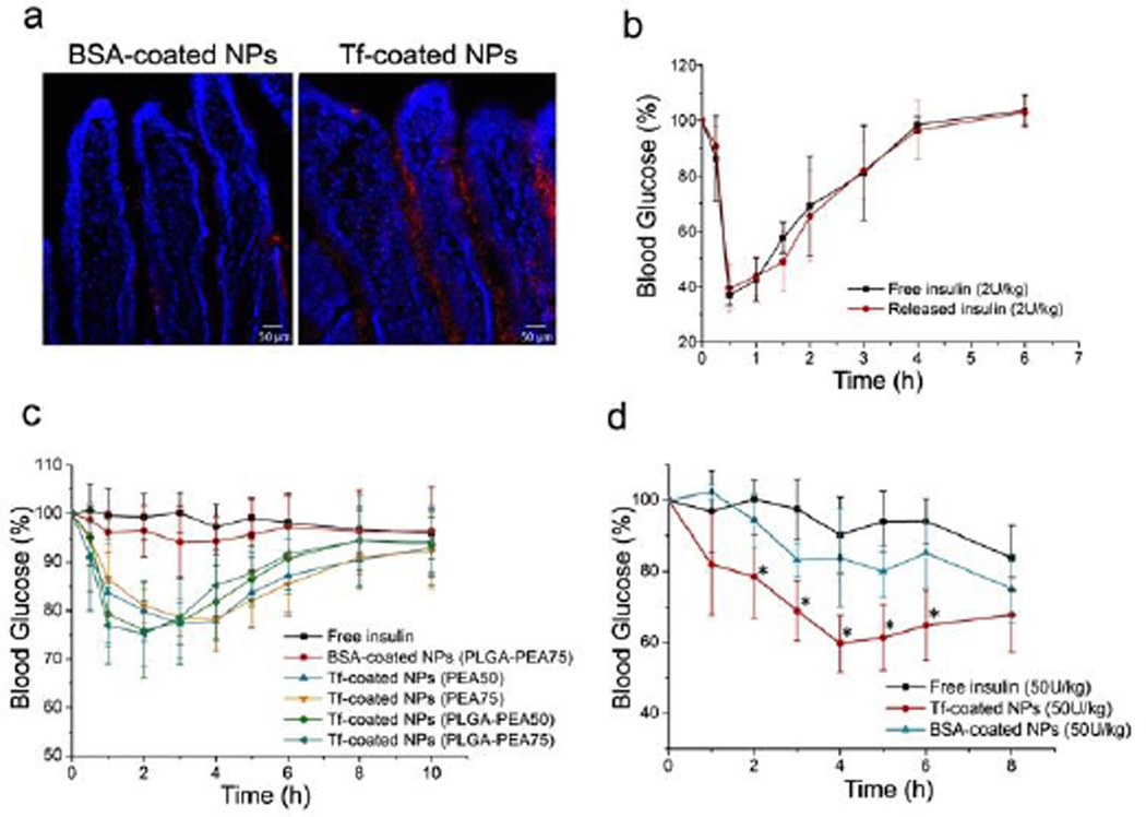
Figure 4.
(a) Fluorescence images of sections of mouse intestine after administration of BSA- or Tf-coated NPs (red). Cell nuclei were stained with DAPI (blue); (b) Blood glucose response of normal rats to free insulin or insulin released from the NPs (2 U/kg) (n = 4); (c) Blood glucose response of normal rats to free insulin solution, BSA-coated NPs, and Tf-coated NPs with different formulations following oral gavage (n = 6); (d) Blood glucose response of diabetic mice to free insulin solution, BSA-coated NPs, and Tf-coated NPs following oral administration (n = 6, * p<0.05 vs. free insulin).

