Figure 3.

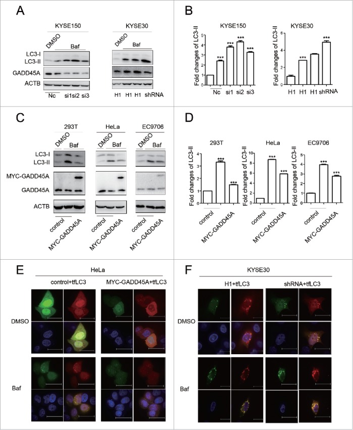
The effect of GADD45A on autophagic flux. (A) KYSE30-H1 and KYSE30-shRNA cells were treated with Baf at 70% to 80% confluence. KYSE150 cells were treated with Baf after GADD45A was knocked down by siRNA. Cell lysates were prepared for analyzing the expression of LC3 and GADD45A. (C) 293T, HeLa and EC9706 cells were treated with Baf after transfection with pCS2-MT or pCS2-MT-GADD45A. LC3 and GADD45A were analyzed using western blots. (B, D) The densities of signals were determined by densitometry and are shown relative to the control group. Graphical data denote mean ± SD. (E, F) HeLa cells were transiently transfected with pCS2-MT, ptfLC3 or pCS2-MT-GADD45A, ptfLC3 vector. Cells were seeded onto the cover slips precoated with polylysine 24 h later after transfection and analyzed by immunofluorescence to detect the expression of RFP and GFP. KYSE30-H1 or KYSE30-shRNA cells were transfected with ptfLC3 vector and seeded onto the cover slips precoated polylysine. Immunofluorescence analyzed the expression of RFP and GFP. Scale bar: 10 μm. ***,P < 0.001.
