Figure 3.
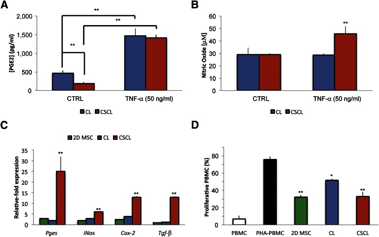
Role of CSCL in supporting MSC immunosuppressive potential in vitro. MSCs secrete various factors, such as PGE2, nitric oxide, and transforming growth factor-β. PGE2 (A) and nitric oxide (B) production by MSCs cultured onto CL and CSCL in response to stimulation with the proinflammatory cytokine TNF-α (50 ng/ml) at 48 hours. Asterisks depict highly significant (∗∗, p < .01) differences compared with cells grown in standard media. (C): Comparison between MSCs grown onto CL and CSCL for the expression of Pges, iNos, Cox-2 and Tgf-β after 48 hours of stimulation. Data are represented as fold-change compared with the expression levels found in the untreated cells (n = 3; ∗∗, p < .01). (D): Effect of MSCs grown in two dimensions or onto CL (CL) or CSCL (CSCL) on the proliferation of stimulated PBMC after 72 hours of coculture. For comparison the percentage of proliferative PBMCs in the presence (PHA-PBMC) and absence (PBMC) of PHA is also reported. Data represent the mean ± SD of three independent experiments. Asterisks depict highly significant (∗∗, p < .01) and significant (∗, p < .05) differences compared with stimulated PBMCs. Abbreviations: 2D MSC, two-dimensional mesenchymal stem cell; CL, collagen scaffold; CSCL, chondroitin sulfate crosslinked onto a collagen-based scaffold; CTRL, control; MSC, mesenchymal stem cell; PBMC, peripheral blood mononuclear cell; PGE2, prostaglandin E2; PHA-PBMC, phytohemagglutinin peripheral blood mononuclear cell; TNF-α, tumor necrosis factor-α.

