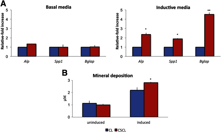
Figure 6.
Osteogenic differentiation in vitro. Mesenchymal stem cells were grown onto CL and CSCL for a 21-day period in basal or inductive media. (A): Quantitative polymerase chain reaction analysis for the osteogenic (Alp, Spp1, Bglap)-associated markers. Expression levels normalized to the reference gene (Gapdh). Data are represented as fold-change compared with expression cells grown on CLs. (B): Mineral deposition (μM) evaluated after 21 days of culture onto CL and CSCL whether cells were exposed to inductive media. Values are mean ± SD. n = 3. Asterisks depict significant (∗, p < .05) and highly significant (∗∗, p < .01) differences between CL and CSCL. Abbreviation: CL, collagen scaffold; CSCL, chondroitin sulfate crosslinked onto a collagen-based scaffold.

