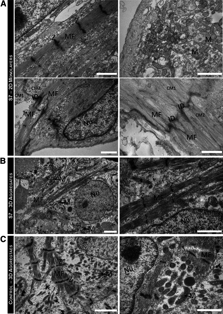
Figure 5.
Ultrastructural characterization of hiPSC-CMs after hypothermic preservation. (A, B): Transmission electron microscopy (TEM) images of hiPSC-CMs, stored at hypothermic conditions for 7 days, as 2D monolayers (A) and as 3D aggregates (B). (C): TEM images of the control 3D aggregates: hiPSC-CMs not subjected to hypothermic storage, that is, maintained in culture during the storage period (control). HiPSC-CMs presented a large nucleus (Nu)-to-cytoplasm ratio and contained many myofibrils aligned and organized in a sarcomeric pattern. Z-bands and neighboring CMs (CM1 and CM2) connected by intercalated disks were also observed. Scale bars = 1 μm (A, B right, C right) and 2 μm (B left, C left). Abbreviations: 2D, two-dimensional; 3D, three-dimensional; CM, cardiomyocyte; hiPSC-CM, human induced pluripotent stem cell-derived cardiomyocyte; ID, intercalated disks; M, mitochondria; MF, myofibrils; Nu, nucleus; S7, stored at hypothermic conditions for 7 days; Z, Z-band.

