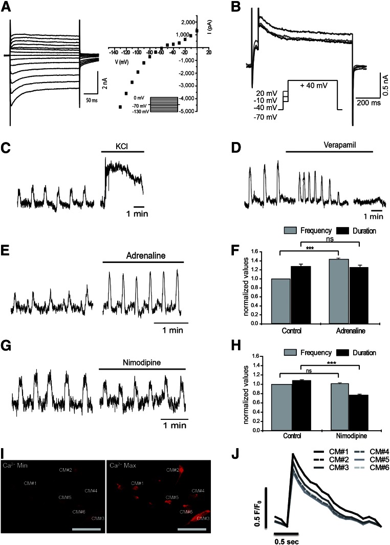
Figure 6.
Functional characterization of hiPSC-CMs subjected to hypothermic storage for 7 days. After hypothermic storage for 7 days and 1 week in culture after storage, cell functionality was evaluated by the detection of CM-specific action potentials (AP) (A, B), typical CM response to drugs (C–H), and calcium transients (I, J). (A): Whole-cell voltage-clamp recordings from hiPSC-CMs subjected to 7 days of hypothermic storage as 2D monolayers. (A): Left: Representative currents following a set of voltage pulses (260 ms), covering a wide range of potentials, with incremental depolarization steps (10 mV) from −130 to 0 mV (holding voltage −70 mV) (inset). Right: Corresponding current-voltage relationship showing three different components in terms of voltage dependence: a strong inward rectifier, an inward hump, and a delayed outward current. (B): Representative current traces from a triple set of double depolarizing pulses: one first step to −40, −10, and +20 mV, lasting 50 ms, followed by a second pulse to +40 mV, lasting 750 ms (inset). One can notice a larger outward current at +40 mV when preceded by a prepulse to −10 mV, which suggest a Ca2+-dependent current component. (C–H): Effect of chronotropic drugs: 30 mM KCL (C), 1 μM Verapamil (D), 0.5 mM Adrenaline (E, F), and 300 pM Nimodipine (G, H), on contractile activity of hiPSC-CMs (cultured as 2D monolayers) subjected to 7 days of hypothermic storage, analyzed by video recording in an inverted microscopy. (I): Pseudocolor images showing minimal (Ca2+ min; left) and maximal (Ca2+ max; right) intensity of the calcium indicator dye Rhod-3. The six hiPSC-CMs analyzed (from plated 3D aggregates) are highlighted. (J): Graphical representation of calcium level cycling, during hiPSC-CMs contraction, determined by confocal imaging of the Rhod-3 dye, for the six different hiPSC-CMs. Data are presented as mean ± SD of 24 (F, H) measurements. ∗∗∗, p < .001; ns, as determined by unpaired t test. Abbreviations: CM, cardiomyocyte; hiPSC-CM, human induced pluripotent stem cell-derived cardiomyocyte; Max, maximal; Min, minimal; ns, not significant.

