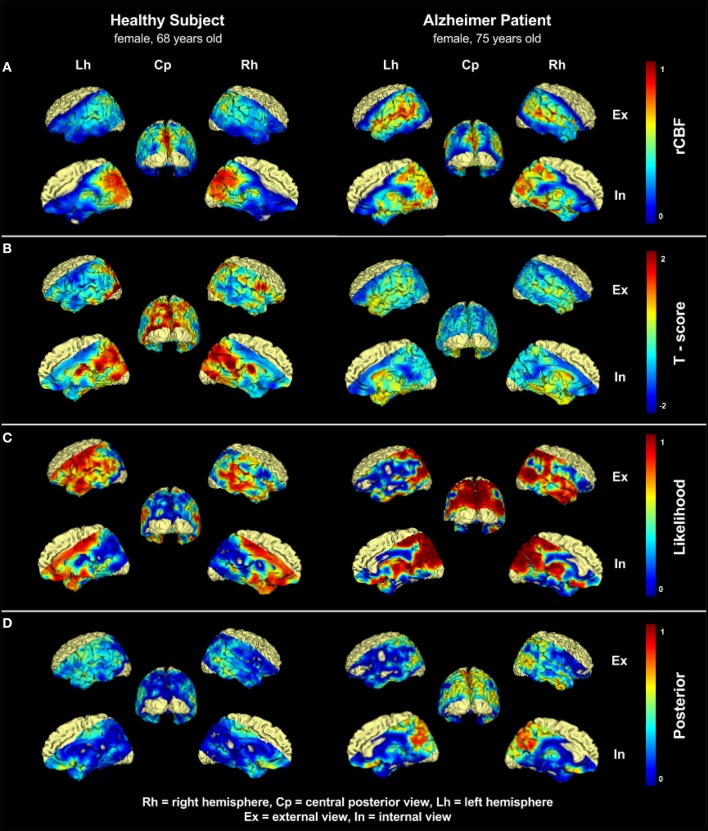
Figure 2.
Comparison of one 75 years old patient with Alzheimers disease (AD) and one 68 years old healthy control (HC). (A) shows the normalized CBF distribution, (B) shows the Tscore-map computed voxel-wise. A substantial difference could be already spotted in the parietal lobe. (C) shows the likelihood of hypo-perfusion L. In the AD patient severely hypo-perfused areas in the parietal lobe and neighboring regions are visible, whereas the HC subject shows regions with artificially low perfusion in the frontotemporal lobes and in the volume boundaries. These high values are the effect of uncertainty resulting from image noise, and/or patient-specific conditions. (D) shows the posterior probability (p), which represents the disease related likelihood of hypo-perfusion resulting from the application of the prior distribution. Areas with artificially low perfusion on volume boundaries have been suppressed, while the the sensitivity to interesting hypo-perfused region in the parietal lobe is still preserved. Upper labels Rh, Cp, and Lh indicates right hemisphere, central posterior, and left hemisphere, respectively. Lateral labels Ex and In indicates external view and internal view, respectively.

