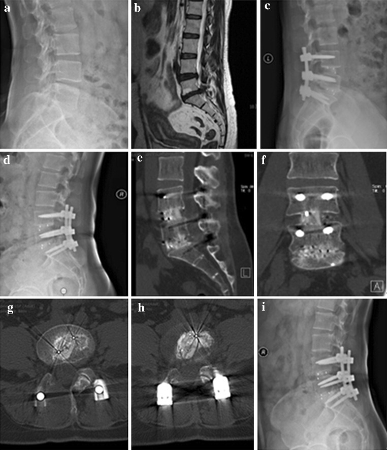
Fig. 4.
A 42-year-old female who underwent 2-level TLIF with n-HA/PA66 cages for lumbar reconstruction. The preoperative lumbar radiographs (a, b). The 1-week postoperative and 3-month follow-up radiographs (c, d). The CT or 3D-CT scan (e, f, g, h) shows that the autogenous bone granules fill the cages and achieve bony fusion with adjacent endplates by the 10-month follow-up. A lateral radiograph (i) at the final follow-up shows satisfactory bony fusion and no obvious migration, radiolucent gap or subsidence

