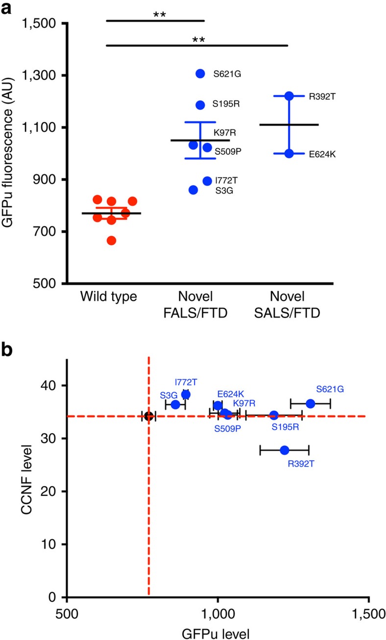
Figure 2. Mutant cyclin F impairs ubiquitin-mediated proteasomal degradation.
NSC-34 cells were co-transfected with GFPu and either wild type or mutant cyclin F, tagged with mCherry. GFPu fluorescence intensity was analysed by flow cytometry 48 h post transfection. (a) Plot of GFPu fluorescence intensity following flow cytometry. A significantly higher level of GFPu fluorescence was observed in cells expressing novel cyclin F mutations (blue data points) when compared with those expressing wt CCNF (red data points) WT v FALS/FTD P=0.0017, d.f.=11; WT v SALS/FTD P=0.001, d.f.=7; two-tailed unpaired Student's t-test). (b) The higher level of GFPu fluorescence was independent of the level of cyclin F as quantified using mCherry signal—R-squared=0.13. Red dashed lines represent the WT mean. Data are represented as mean,±s.e.m. n=3 (n is one experiment consisting of the mean of 50,000 cells); **P<0.01. d.f., degrees of freedom.

