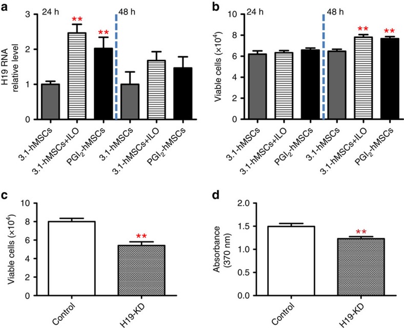
Figure 7. H19 lncRNA promoted host myoblasts survival and proliferation under hypoxia (1.5% O2).
(a) Primary myoblasts co-cultured with PGI2-hMSCs or with 3.1-hMSCs+ILO for 24 h showed a significant increase in H19 lncRNA levels as compared with those co-cultured with 3.1-hMSCs. The increase in H19 lncRNA levels was transient; at 48 h, levels were comparable among all the groups. (b) No differences were seen in the total number of viable cells among myoblasts co-cultured with PGI2-hMSCs, 3.1-hMSCs+ILO or 3.1-hMSCs for 24 h. However, by 48 h, the number of viable myoblasts was significantly higher when cells were co-cultured with PGI2-hMSCs or 3.1-hMSCs+ILO than with 3.1-hMSCs. (c) H19 silencing reduced the paracrine effects of PGI2-hMSCs on myoblast survival. The total number of viable cells was significantly reduced in myoblasts with H19 deficiency (H19 KD) that were co-cultured with PGI2-hMSCs for 48 h as compared with control myoblasts co-cultured with PGI2-hMSCs in parallel. (d) The paracrine effects of PGI2-hMSCs on myoblast proliferation were also impaired with H19 silencing. Myoblasts with H19 deficiency that were co-cultured with PGI2-hMSCs for 48 h showed a significant reduction in cell proliferation as compared with control myoblasts co-cultured in parallel with PGI2-hMSCs. **P<0.01. Statistical significance was determined by one-way ANOVA with Newman–Keuls post hoc test (a,b) and a two-tailed t-test (c,d). Data are shown as mean±s.e.m. from three to four replicates and are representative of three independent experiments with similar results.

