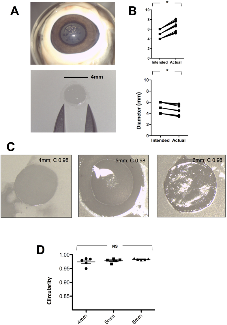
Figure 1. Capsule retrieval, time to completion and circularity.
Color photographs showing the capsulotomy and fragmentation pattern in situ, retrieval, measurement and mounting of lens capsule prior to preparation for scanning electron microscopy (A). The intended and actual size of capsulotomy (upper panel) and capsule (lower panel) (B). Representative photographs of lens capsule circularity at 4, 5 and 6 mm (n = 18) (C) and showing comparison at different sizes (n = 15) (D). **p < 0.01, NS = Not Significant

