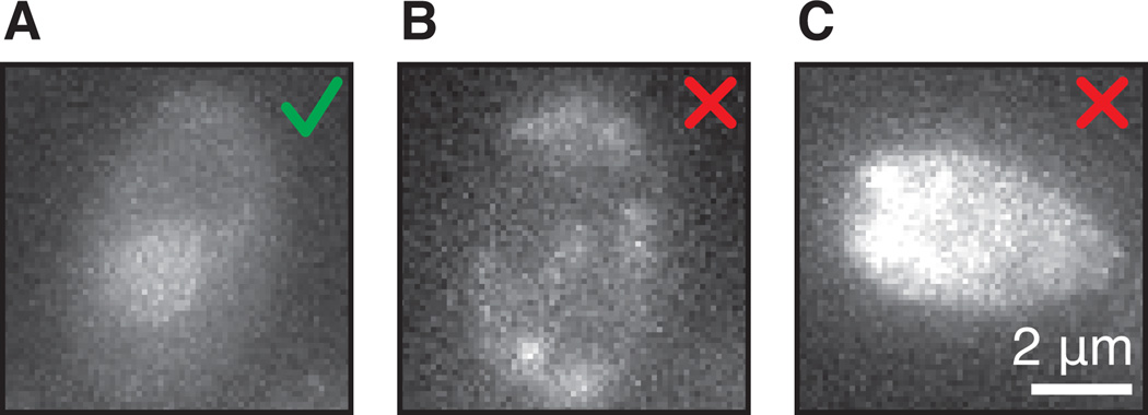
Figure 3.
(A) Example of a correct coat protein levels in the cell. There is even staining across the cell and slightly higher coat protein levels in the nucleus. (B) Example of photo toxicity. The cell shows uneven fluorescence at the cell membrane. (C) Example of auto-fluorescent cell grown into diauxic shift.

