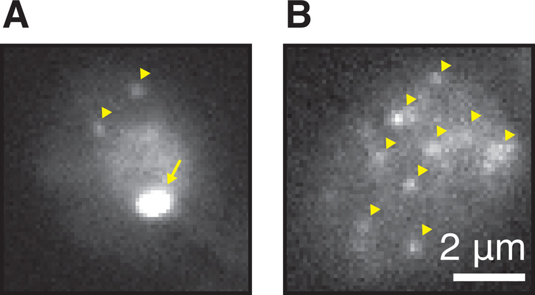
Figure 5.
Detection of single mRNAs. (A) GAL10 PP7 tagged cell in galactose imaged with fast exposure 30ms. Note the nuclear staining, the bright transcription site (arrow) in the nucleus and two cytoplasmic RNAs (arrowheads). Frequently, the transcription site will appear saturated at light intensities where single mRNAs are visible. (B) Same as panel A with several single RNAs (arrowheads). Note that the nucleus is out of focus.

