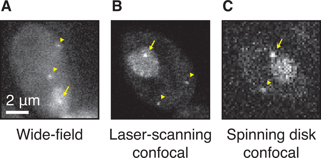
Figure 6.
Detection of single RNAs with different microscopes. (A) Single RNAs tagged with PP7, imaged with a wide-field or epifluorescence microscope, 50ms exposure. The transcription site is marked with an arrow, the diffusing RNAs with arrowheads. (B) Same strain as panel A, imaged with a laser-scanning microscope. (C) Same as panel A, image with a spinning disk confocal microscope, 50ms exposure.

