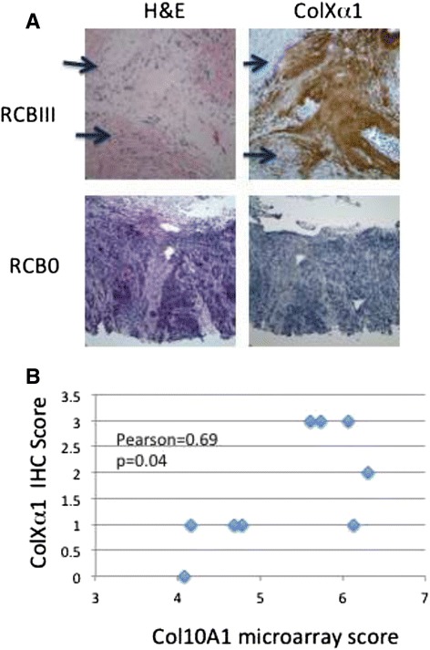
Fig. 3.

Immunohistochemistry of colXα1. a Representative colXα1 immunostaining in low- and high- colXα1 expressing ER+/HER2+ breast cancers. Two representative cases, one with no response, RCBIII, and strong colXα1 signal, score = 2, and one with good response, RCB0, and no colXα1 signal, score = 0, are shown. Arrows indicate regions with tumor cells. b RNA levels as determined by the microarray correlate with colXα1 IHC signal in 9 cases
