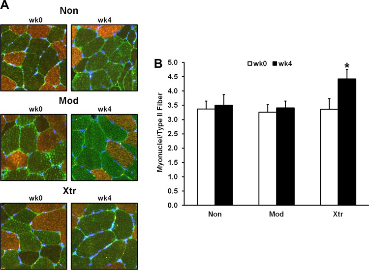
Fig. 4.
Cluster differences in type II myofiber myonuclear addition from pre- to post-RT. A: representative ×20 immunohistochemical images [nuclei labeled with 4′,6-diamidino-2-phenylindole (DAPI)]. B: average no. of myonuclei per type II myofiber within each responder cluser before resistance training (wk0) and posttraining (wk4). *Significantly different from previous time point (P < 0.05). Non, nonresponder; Mod, modest responder; Xtr, extreme responder.

