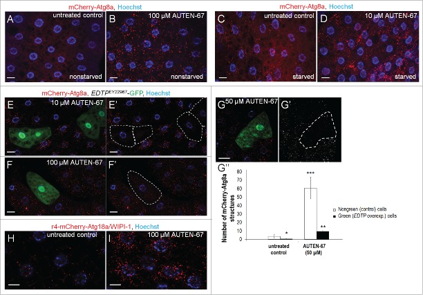
Figure 3.
AUTEN-67 induces autophagy in Drosophila via inhibiting EDTP. (A) Fat body cells from a feeding L3 stage larva (90 h) transgenic for a mCherry-Atg8a reporter show basal levels of autophagic activity. (B) AUTEN-67 (100 µM) treatment results in a massive accumulation of Atg8a-positive structures in the fat body of a L3F larva. (C) Fat body cells of a starved L3 stage larva (90 h) accumulate autophagic structures. (D) AUTEN-67 (10 µM) increases the amount of Atg8a-positive structures in fat body cells of a starved L3 larva, as compared to untreated control. (E to G”) Clonal overexpression of EDTP in fat body cells largely, but not completely, inhibits the formation of autophagic structures (red foci) induced by AUTEN-67 at 10 µM (E and E'), 100 µM (F and F') and 50 µM (G and G') concentrations. EDTP-overexpressing cells are green in the left panels, and outlined by dotted lines in the corresponding right panels. (G”) Quantification of autophagic structures in Drosophila fat body cells treated with AUTEN-67 (50 µM). EDTP-overexpressing (green/outlined) cells contain much fewer autophagic structures than nongreen controls (*: P <0.05, **: P<0.01, ***: P<0.001; paired Student t test). Bars represent s.e. (H) Expression of Atg18a/WIPI1 in fat body cells from a untreated L3 stage larva (control). Red foci label phagophores. (I) Atg18a/WIPI1 abundantly accumulates in fat body cells of an L3 stage larva treated with 100 uM AUTEN-67. In figures A to G, H and I, bars indicate 10 µm, blue coloring (Hoechst staining) labels nuclei.

