Figure 1.

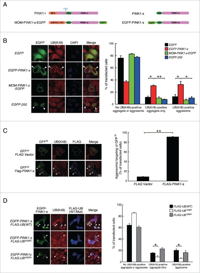
Overexpression of PINK1-s induces the formation of aggregates and aggresomes enriched with K48-linked UB chains. (A) Schematic illustration of the EGFP-tagged PINK1 proteins mimicking the endogenous cytosolic PINK1-s and mitochondrial PINK1-l. A 67-amino acid peptide from the mitochondrial outer membrane (MOM) protein USP30 was fused to the N terminus of PINK1-s-EGFP to target it to the MOM, mimicking the PINK1-l accumulation on the outer membrane of damaged mitochondria. The PARL cleavage site is indicated by arrowheads. (B) Aggregate and aggresome formation in AD293 cells transiently overexpressing EGFP, EGFP-PINK1-s, MOM-PINK1-s-EGFP or EGFP-250 for 24 h. UB-containing aggregates and aggresomes were visualized by immunofluorescence staining with a UB antibody specific for K48-linked chain (UB(K48) antibody). Aggresomes are indicated by arrowheads. Scale bar: 20 μm. Quantification results are shown as mean ± SEM of 3 independent experiments. *, P < 0.05; **, P < 0.01. (C) Aggresome formation in AD293 cells transiently expressing GFPU (Control) or GFPU and FLAG-PINK1-s for 24 h. GFPU, ubiquitinated proteins and FLAG-PINK1-s were detected by immunofluorescence staining with EGFP, UB(K48) and FLAG antibodies, respectively. Aggresomes are indicated by arrowheads. Scale bar: 20 μm. Qualification results are shown as mean ± SEM of 3 independent experiments. **, P < 0.01. (D) Aggresome formation in AD293 cells transiently expressing EGFP-PINK1-s together with FLAG-UB(WT), FLAG-UBK48R or FLAG-UBK63R for 24 h. EGFP-PINK1-s, K48-linked ubiquitin chain, FLAG-UB were detected by immunofluorescence staining with EGFP, UB(K48) and FLAG antibodies, respectively. Aggresomes are indicated by arrowheads. Scale bar: 20 μm. Qualification results are shown as mean ± SEM of 3 independent experiments. *, P < 0.05.
