
Figure 7.
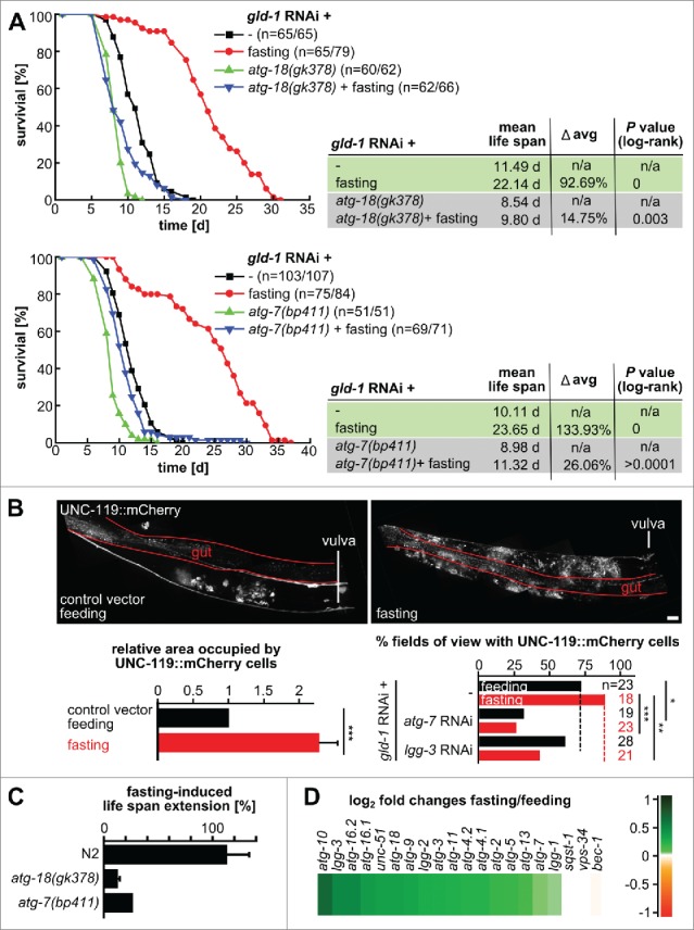
Fasting delays death caused by germline tumors in an autophagy-dependent manner and promotes neuronal differentiation. N2, atg-18 and atg-7 mutant L1 larvae were fed with gld-1 RNAi till the d 2 of adulthood and then they were either fed with bacteria expressing empty vector or fasted throughout their life span. (A) Representative cumulative survival curves and Kaplan Meier statistics are presented. (B) Top: UNC-119::mCherry L1 larvae were treated with gld-1 dsRNA till d 2 of adulthood and then treated as indicated for 1 d. Representative gonad arm reconstructed from maximum projections. Scale bar: 20 μm. Bottom left: Area occupied by UNC-119 expressing cells per gonad arm normalized by the animals kept under feeding conditions. Qualitative scoring of the gonadal area occupied by UNC-119 expressing cells was performed blindly using maximum z-projections covering whole gonad arms (d 3 of adulthood). Data represent mean ± SEM of 2 independent experiments, n = 5 (10 gonad arms), per experiment; *** P ≤ 0.001. Bottom right: Quantification of tumors with UNC-119-expressing cells. Animals were treated with the RNAis as indicated and either continued feeding or were starved for 1d before imaging and scoring. (C) Fasting-induced life span of gld-1 RNAi-treated N2, atg-18 and atg-7 mutants. For N2 and atg-18 mutant, data represent mean ± SEM of 2 independent experiments. (D) Expression profiles of transcripts in the autophagy pathway of fasted gld-1 RNAi animals compared to fed gld-1 RNAi animals. log2-fold changes are shown.