
Figure 1.
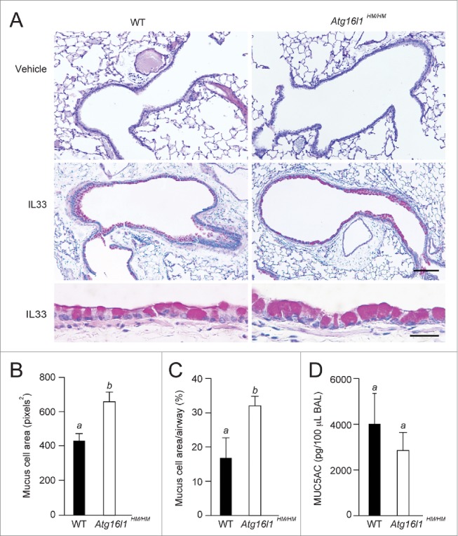
Goblet cell hypertrophy in autophagy-deficient mice. WT and Atg16l1 hypomorphic (Atg16l1HM/HM) mice were administered vehicle or IL33 intranasally. Lungs were evaluated on d 7 post-challenge. (A) Representative photomicrographs of lung sections stained with PAS. Scale bars, top and middle panels: 100 μm; lower panel: 10 μm. (B) Quantification of area per goblet cell (n = 6 mice per group, (C) percentage of PAS staining (on histological sections) of total airway epithelium area (n = 6 mice per group) of IL33-treated WT and Atg16l1HM/HM, and (D) levels of MUC5AC in bronchoalveolar lavage (BAL) fluid from lungs of IL33-treated mice (n = 3 mice per group). (B–D) are graphs of the means ±SEM. Means with different letters are significantly different by the unpaired 2-tailed Student's t-test.