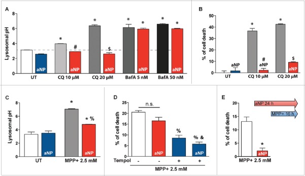
Figure 3.
Acidic nanoparticles rescued lysosomal pH after lysosomal inhibitor treatment and PD-related toxin exposure. (A) Lysosomal pH values as measured ratiometrically using LysoSensor Yellow/Blue DND-160 in untreated cells, 10 and 20 µM chloroquine (CQ) or 5 and 50 nM bafilomycin A1 (BafA)-treated cells, with or without PLGA-aNP incubated concurrently for 24 h or 1 h respectively. (B) Cell death in CQ-treated cells incubated with or without PLGA-aNP. (C) Lysosomal pH values in untreated (UT) and MPP+-intoxicated M17 cells, in the absence or presence of PLGA-aNP treatment. (D) Cell death in MPP+-treated M17 cells after incubation with PLGA-aNP for 24 h, in the presence or the absence of Tempol (500 µM). (E) Cell death measured after PLGA-aNP incubation for 24 h prior to 16 h of MPP+ treatment. In all panels, n=3 to 5 per experimental group. *, P<0.05 compared with control untreated cells; #, P<0.05 compared with CQ (10 µM)-treated cells; $, P<0.05 compared with CQ (20 µM)-treated cells; %, P<0.05 compared with MPP+-treated cells; and &, P<0.05 compared with MPP+-intoxicated cells treated with PLGA-aNP.

