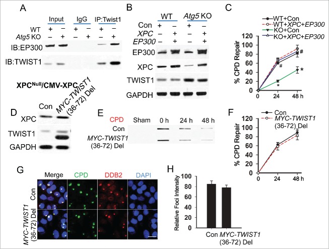
Figure 5.
Autophagy deficiency inhibits DDB2 recruitment through Twist1 binding and inhibition of EP300. (A) Immunoprecipitation was performed in WT and atg5 KO MEF cells with the indicated antibodies, followed by immunoblot analysis of EP300 and TWIST1. (B) Immunoblot analysis of EP300, XPC, TWIST1 and GAPDH in WT and atg5 KO MEF cells transfected with Con or the combination of Xpc and Ep300. (C) Quantification of percentage (%) of CPD repair in WT and atg5 KO MEF cells transfected with Con or the combination of Xpc and Ep300 (mean±SD, n=3). *, P < 0.05, compared with WT group; #, P < 0.05, compared with Con group, Student t test. (D) Immunoblot analysis of XPC, TWIST1 and GAPDH in XPC-/--CMV-XPC cells transfected with Con and MYC-TWIST1 (36 to 72) deletion. (E) Slot blot analysis of the levels of CPD in XPC-/--CMV-XPC cells transfected with Con and MYC-TWIST1 (36 to 72) deletion. (F) Quantification of percentage (%) of CPD repair from (E) (mean±SD, n=3). *, P < 0.05, compared with WT group; #, P < 0.05, compared with Con group, Student t test. (G) Immunofluorescence assay of the colocalization of DDB2 with subnuclear CPD in HaCaT cells transfected with Con or MYC-TWIST1 (36 to 72) deletion at 0.5 h post-UV (10 mJ/cm2) through a 5-μm micropore filter. Scale bar: 10 μm. (H) The relative intensity of DDB2 was calculated by analyzing 100 foci and normalized to that of CPD (n =100, Mean±SD). All results were obtained from 3 independent experiments.

