
Figure 6.
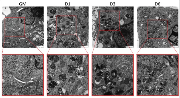
Electron micrographs of differentiating C2C12s treated with BAF. Transmission electron microscopy was performed on differentiating C2C12s treated with 100 nM BAF to examine alterations in mitochondrial populations. Insets are presented at higher magnification below each original image. Scale bars: 500 nm. GM, growth medium.