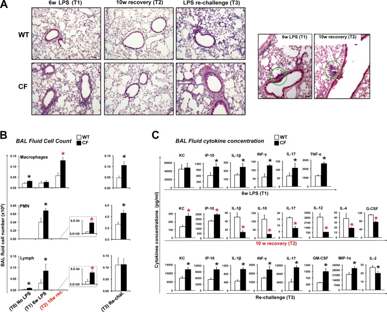
Fig. 3.
Chronic LPS exposure leads to unresolved lung inflammation in CF mice. A: representative hematoxylin-eosin staining of paraffin-embedded lung tissues from WT and CF mice at T1, T2, and T3. Representative images showing lung lymphocyte aggregates (T1 and T2), which were similarly distributed in both genotypes, are shown at left. Dense lymphocytic aggregation is shown by green circle. Scale bars = 20 μm. B and C: differential bronchoalveolar lavage (BAL) fluid cell number and BAL fluid cytokine concentration in WT and CF mice at T1, T2, and T3. Note difference in scale bars in B, right and left. KC, keratinocyte chemoattractant; IP-10, IFN-γ-induced protein 10; G-CSF, granulocyte colony-stimulating factor; GM-CSF, granulocyte-macrophage colony-stimulating factor; MIP-1α, macrophage inflammatory protein-1α. Values are means ± SE. *P < 0.05.

