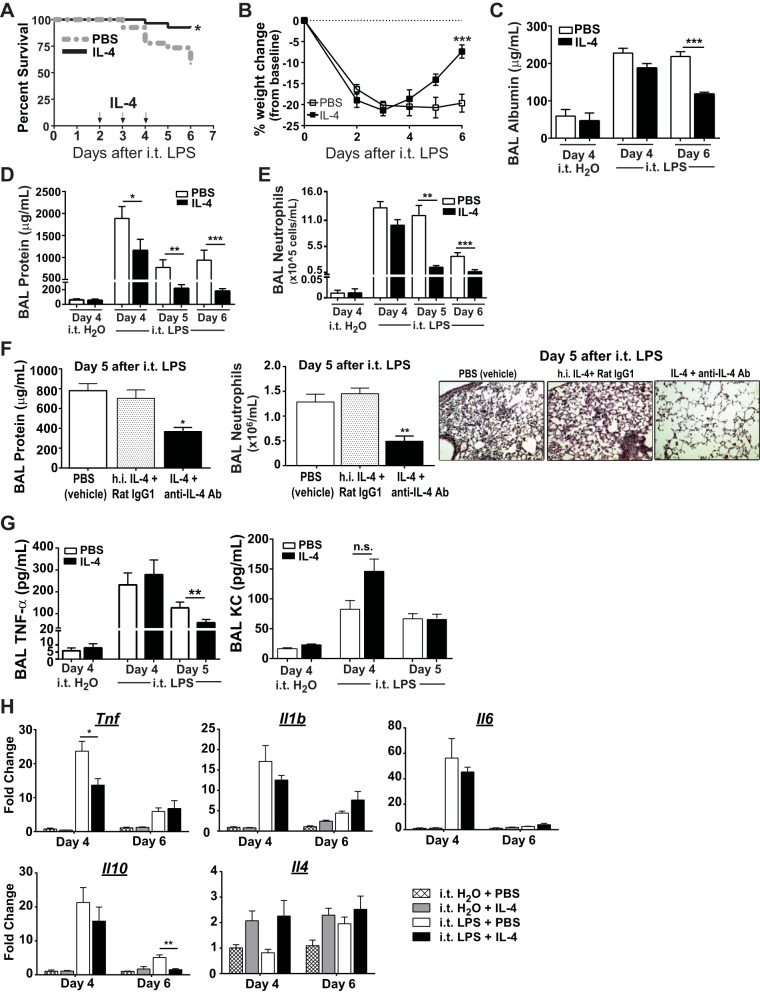
Fig. 1.
Wild-type (WT; C57BL/6) mice treated with IL-4 demonstrate improved survival and accelerated acute lung injury (ALI) resolution. A: Kaplan-Meier survival curves for mice exposed to intratracheal (i.t.) LPS (4 mg/kg) and treated with IL-4 or PBS (n = 27–30, P = 0.006 by Mantel-Cox). B: daily percent weight change from baseline for each group following intratracheal LPS (n = 6–10, ***P < 0.001 by repeated-measures ANOVA). Bronchoalveolar lavage (BAL) albumin (C) and BAL protein (D) values, as well as BAL neutrophil (E) numbers at day 4 in intratracheal H2O-exposed mice, or at days 4–6 in intratracheal LPS-exposed mice (n = 4 for intratracheal H2O groups, 6–10 for intratracheal LPS groups, *P < 0.05, **P < 0.01 by one-way ANOVA). F: following intratracheal LPS, mice received either PBS (vehicle), heat-inactivated (h.i.) IL-4 + Rat IgG1 (protein control), or IL-4 + anti-IL-4 Ab (active IL-4) on days 2–4. BAL protein and BAL neutrophils were quantified at day 5 after intratracheal LPS (n = 7 per group, *P < 0.05, **P < 0.01 by one-way ANOVA). Representative hematoxylin and eosin stain of the lung at day 5 after intratracheal LPS in all 3 groups at ×20 magnification. G: BAL TNF-α and KC after intratracheal H2O or LPS (**P < 0.01 by t-test, n = 4 for intratracheal H2O groups, 5–6 for LPS groups). H: gene expression changes for Tnf, Il1b, Il6, Il10, and Il4 in lung tissues following intratracheal LPS or H2O in IL-4- or PBS-treated mice (n = 3–6, *P < 0.05, **P < 0.01 by one-way ANOVA).

