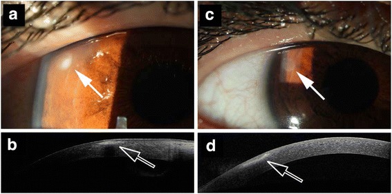
Fig. 1.

Pre- and post-treatment peripheral infectious keratitis. a Anterior segment photography of a patient with early peripheral infectious keratitis (arrow). b Anterior segment OCT of the lesion. c Anterior segment photography of the same patient 7 days after PACK-CXL (9 mW/cm2 irradiance for 10 min) with resolution of the peripheral infectious keratitis (arrow). d Anterior segment OCT of the lesion at day 7 after PACK-CXL
