Fig. 3.

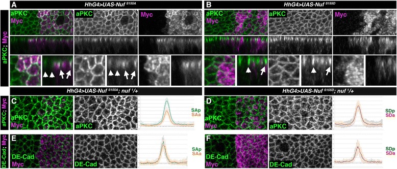
NufS155A increases aPKC apico-lateral membrane levels. a-b Wing discs of wild-type larvae expressing in the posterior compartment Myc-NufS155A (a) or Myc-NufS155D (b) stained for aPKC (green) or Nuf (anti-Myc, magenta). Upper panels apical views, medial panels sagittal views and lower panels close-up of the above. Arrowheads and arrows point to cortical aPKC in cells located in anterior or posterior compartment of disc. c-f Quantification of aPKC (c-d) and DE-Cad (e-f) levels in epithelia of nuf 1/+ heterozygous background wing disc expressing in the posterior Myc-NufS155A (c, e) or Myc-NufS155D (d, f). Fluorescence levels of aPKC or DE-Cad are shown comparing control anterior cells (orange and red) with posterior cells expressing Myc-NufS155A or Myc-NufS155D (blue). Nuf nuclear fallout, aPKC atypical PKC
