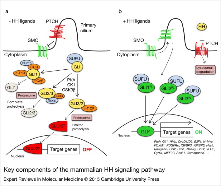
Figure 1.
Key components of the mammalian HH signalling pathway. In absence of HH ligands (a), PTCH inhibits SMO by preventing its entry into the primary cilium. GLI proteins are phosphorylated by PKA, GSK3β and CK1, which create binding sites for the E3 ubiquitin ligase β-TrCP. GLI3 and, to a lesser extent, GLI2 undergo partial proteasome degradation, leading to the formation of repressor forms (GLI3/2R, red), that translocate into the nucleus where they inhibit the transcription of HH target genes. Full-length GLI may also be completely degraded by the proteasome. This process can be mediated by Spop and Cullin 3-based E3 ligase for GLI2 and GLI3, whereas GLI1 can be degraded by β-TrCP, the Numb-activated Itch E3 ubiquitin ligase and by PCAF (see text for details). Upon HH ligand binding (b), PTCH is displaced from the primary cilium, allowing accumulation and activation of SMO. Active SMO promotes a signalling cascade that ultimately leads to translocation of full length (FL) activated forms of GLI (GLIA, green) into the nucleus, where they induce transcription of HH target genes. Abbreviations: CK1, casein kinase 1; GSK3β, glycogen synthase kinase 3β; HH, Hedgehog; PCAF, p300/CREB-binding protein (CBP)-associated factor; PKA, protein kinase A; PTCH, Patched; SMO, Smoothened; Spop, speckle-type POZ protein; SUFU, Suppressor of Fused; β-TrCP, β-transducin repeat-containing protein.

