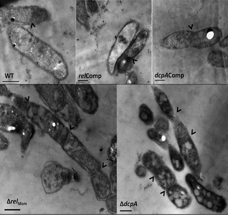
FIG 6.
Ultrastructural analysis of the WT, knockout, and complemented strains using transmission electron microscopy. It can be noted from the transmission electron micrographs that the elongated ΔrelMsm and ΔdcpA cells are multiseptate, unlike the WT and the respective complemented strain. Arrowheads represent the positions of septa. Bars, 500 nm.

