FIG 3.
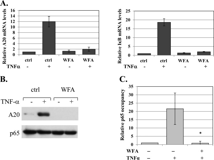
WFA inhibits NF-κB activity in cells. (A) Untreated or WFA-treated (10 μM; 1 h) cells were induced by TNF-α for 1 h, and levels of A20, IκBα, and GAPDH (glyceraldehyde-3-phosphate dehydrogenase) mRNAs were determined by reverse transcription (RT)-qPCR using gene-specific primers. The bars represent the means and standard deviations (SD) of A20 and IκBα levels, normalized to GAPDH, of 2 independent experiments. (B) Untreated or WFA-treated cells (10 μM; 1 h) were induced by TNF-α for 2 h, and the level of A20 protein was determined by Western blotting. The WFA lanes were spliced to bring them close to the control lanes. The original gel is shown in Fig. S4 in the supplemental material. (C) Cells, pretreated with WFA for 60 min and then treated with TNF-α for 30 min or left untreated, were subjected to ChIP using anti-p65 or control (for background levels) antibodies. Analysis was performed by qPCR. The graphs show occupancy levels normalized to the input levels. The uninduced sample was set to 1. The results represent the averages ± SEM of the results of 3 independent experiments. The asterisk denotes a statistically significance difference (P < 0.05).

