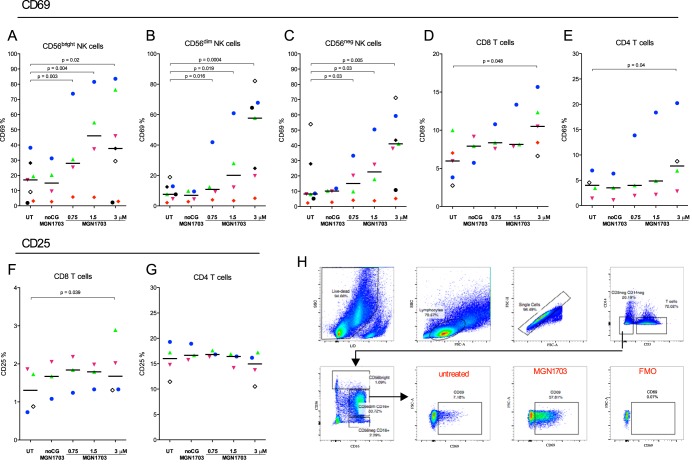
FIG 2.
MGN1703 activates NK cells. PBMCs from ART-suppressed HIV-1-infected donors were incubated for 48 h with MGN1703 at the indicated concentrations. Controls included noCG-MGN1703 as a TLR9-specific negative control or cRPMI as an untreated control. Cells were stained and then assessed by flow cytometry. (A to C) The median percentage of CD69-positive CD56dim (CD56dim CD16+) NK cells increased up to 7.5-fold in response to MGN1703. Also, CD56bright (CD56bright CD16+/−) and CD56neg (CD56neg CD16+) NK cells increased the proportion of CD69-positive cells by 2.2- and 5-fold, respectively (for comparative results for CpG-ODN2006, see Fig. S2 in the supplemental material). (D and E) The proportions of activated CD8+ and CD4+ T cells were significantly increased. (F and G) Marginally more CD8+ T cells were CD25 positive, while the proportion of CD25-positive CD4+ T cells did not increase. (H) Representative flow diagram for one donor (represented by green triangles), showing the gating strategy for CD69 expression on CD56dim NK cells. Each donor in Fig. 1 to 4 is represented by the same distinct symbol. Lines represent the median. SSC, side scatter; FSC, forward scatter.

