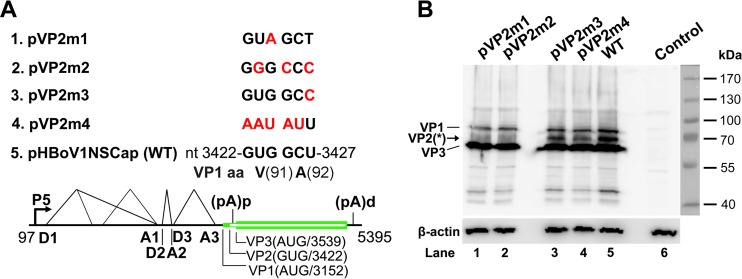
FIG 3.
VP2 is translated from a noncanonical translation initiation codon. (A) Diagrams of pHBoV1NSCap-based mutants. The parent pHBoV1NSCap plasmid is diagrammed with transcription, splicing, and polyadenylation units shown, together with the mutants that carry various mutations at the GUG and GCU codons (nt 3422 to 3427) of the HBoV1 genome, as indicated (red). (B) Western blot analysis of capsid proteins. HEK 293 cells were transfected with plasmids as indicated. The lysates of the transfected cells were analyzed by Western blotting using an anti-VP antibody and reprobed with anti-β-actin. The identities of the detected bands are indicated at the left of the blot. The arrow shows the novel VP2 protein, which was denoted previously by an asterisk (17). Control, without transfection.

