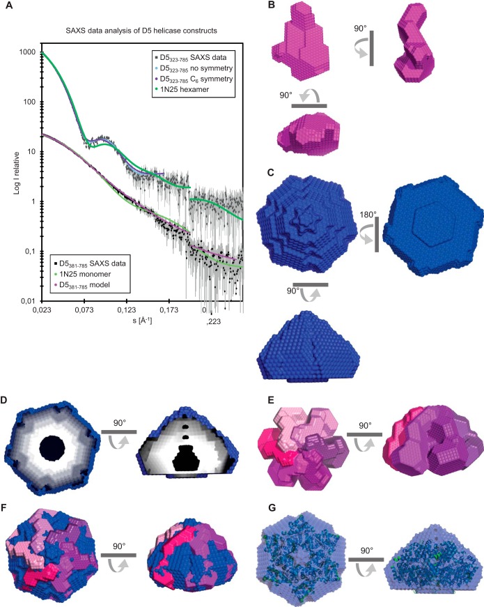
FIG 3.
Models of the helicase domain containing fragments D5381–785 and D5323–735 obtained from SAXS. (A) SAXS scattered intensity (I) of D5381–785 and D5323–735 and calculated intensity as a function of the magnitude of the scattering vector (s) for the models of both proteins with or without the use of C6 symmetry. The scattering curve calculated for SV40 Lta (PDB accession number 1N25) is also shown. Intensities are on an arbitrary scale. (B) Views of the bead model of a D5381–785 monomer (magenta). (C) Model of the D5323–735 hexamer (blue). (D) Cuts through the hexamer model showing the internal cavity. (E, F) Assembly of 6 monomeric models into a hexameric model of D5323–735 (E) and a comparison with the model of the hexamer shown in panel C (F). (G) Overlay of the models of hexameric D5323–735 (blue) and SV40 Lta (cartoon representation in green).

