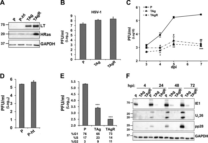
FIG 1.
The expression of SV40 TAg blocks HCMV replication. (A) The protein levels of SV40 large T antigen (LT) and H-Ras in primary fibroblasts (P) or their transduced derivatives were measured by Western blotting. These fibroblast lines include those expressing telomerase alone (P-ht), those expressing telomerase plus the SV40 early region (TAg), or those expressing the combination of telomerase, the SV40 early region, and H-RasV12 (TAgR). (B to D) The cells for which the results are shown in panel A were infected with herpes simplex virus (B) or HCMV AD169 (C and D) at an MOI of 3.0. The production of infectious virions at 1 dpi (B), 3 to 7 dpi (C), and 5 dpi (D) was measured by plaque assay. (C) Asterisks indicate significant differences between the values for primary cells and those for TAg-expressing cells (*, P < 0.05; **, P < 0.01; ****, P < 0.001). Number signs indicate significant differences between the values for primary cells and those for TAgR cells (#, P < 0.05; ##, P < 0.01). (E) The cells for which the results are shown in panel A were maintained in serum-free medium for 96 h and infected with HCMV AD169 at an MOI of 3.0. Cell cycle analysis was performed by analyzing the cellular DNA content by flow cytometry at 24 hpi. Asterisks indicate significant differences between the values for primary cells and those for TAg-expressing cells (P < 0.001). (F) The cells for which the results are shown in panel A were infected with HCMV (AD169, MOI = 3.0), and the protein levels of IE1, UL26, pp28, and GAPDH were measured by Western blotting at 4, 24, 48, and 72 hpi. The numbers to the left of the gels in panels A and F are molecular masses (in kilodaltons).

