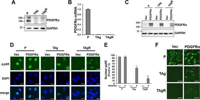
FIG 3.
PDGFRα overexpression partly rescues HCMV entry in fibroblasts expressing SV40 TAg. (A and B) The PDGFRα protein abundance (A) or mRNA level (B) in primary fibroblasts (P) or TAg or TAgR fibroblasts was measured by Western blot and qPCR, respectively. (C) Cell lysates from primary fibroblasts or TAg or TAgR fibroblasts transduced with either PDGFRα or a control vector (Vec) were subjected to Western blotting analysis of PDGFRα. (D) The localization of pp65 in primary fibroblasts or TAg or TAgR fibroblasts transduced with PDGFRα or a control vector was determined by measurement of the level of immunofluorescence at 4 hpi (AD169, MOI = 3.0). Bars, 20 μm. (E) The number of nuclei with pp65 staining in cells infected as described in the legend to panel D was quantified. (F) The indicated cells were infected with EGFP-tagged AD169 (MOI = 3.0), and EGFP fluorescence was measured at 48 hpi. Bars, 100 μm. The numbers to the left of the gels in panels A and C are molecular masses (in kilodaltons).

