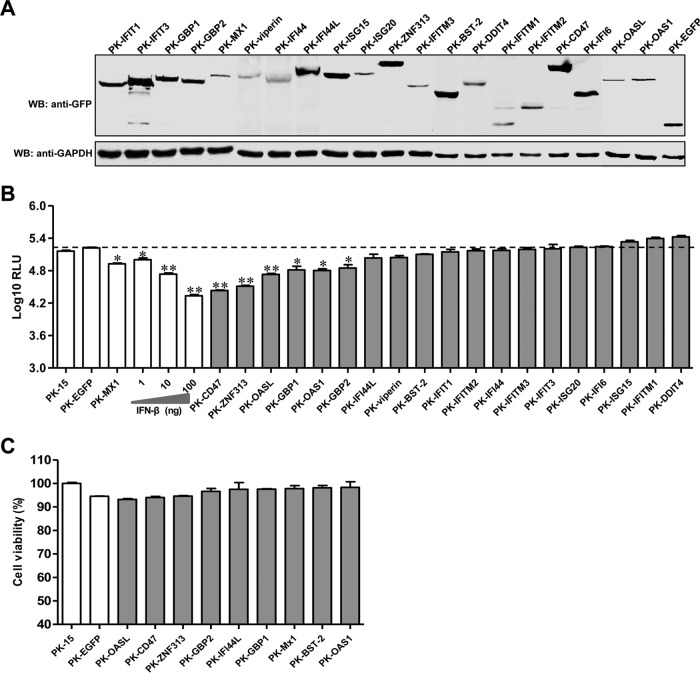
FIG 1.
Screening of ISGs for the ability to inhibit CSFV infection. (A) Characterization of ISG expression in PK-ISG cells. Lysates of PK-ISG or PK-EGFP cells were analyzed by Western blotting (WB) using a mouse anti-GFP (1:1,000) or anti-GAPDH (1:1,000) antibody. (B) Effects of ISG expression on rCSFV-Fluc infection. PK-ISG and PK-EGFP cells were seeded into 48-well plates at a density of 2 × 10⁵ per well. At 24 h postseeding, cells were infected with rCSFV-Fluc at a multiplicity of infection of 0.1, cultured for an additional 48 h, and assayed for luciferase activity using the luciferase reporter assay system (Promega). RLU, relative light units. As controls, parental PK-15 cells were either left untreated or pretreated with the indicated concentrations of IFN-β for 24 h, infected with rCSFV-Fluc, and assayed for luciferase activity at 48 h postinfection as described above. An RLU below the dashed line indicates that the candidate is a potential anti-CSFV ISG. Error bars represent standard deviations. Each sample was run in triplicate.*, P < 0.05; **, P < 0.01. (C) A cell viability assay was performed on cell lines stably overexpressing ISGs.

