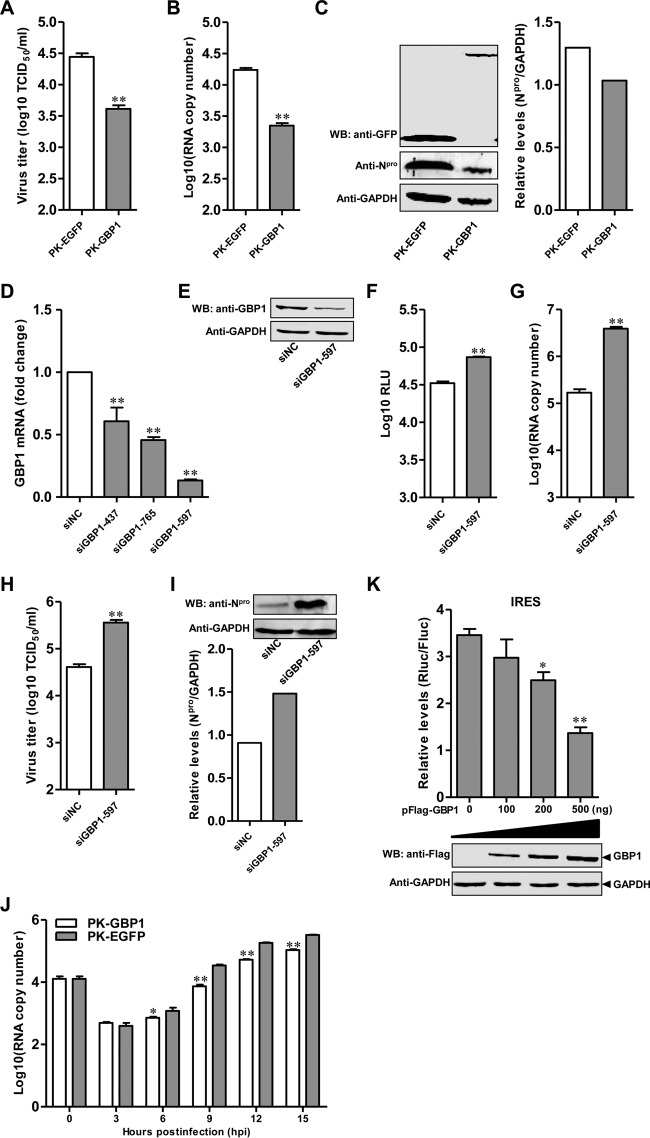
FIG 2.
GBP1 inhibits CSFV replication. (A to C) Influence of GBP1 overexpression on Shimen replication. PK-GBP1 and PK-EGFP cells were infected with CSFV strain Shimen at a multiplicity of infection of 0.1 for 48 h. (A) Virus titers in the supernatants were detected by an immunofluorescence assay and are presented as median tissue culture infective doses (TCID50) per milliliter. Error bars represent standard deviations. *, P < 0.05; **, P < 0.01. (B) The genomic copies of CSFV were assessed using a quantitative real-time reverse transcription-PCR assay. (C) (Left) The expression of Npro in cell lysates was analyzed by Western blotting (WB) using a rabbit anti-Npro polyclonal antibody (1:500). GAPDH protein was used as a loading control. (Right) Quantitative analysis of Npro expression in cell lysates was carried out using Odyssey application software, version 3.0. Each sample was run in triplicate. (D and E) Efficiency of knockdown of GBP1 by siRNAs. (D) PK-15 cells transfected with siGBP1 targeting different sequences (siGBP1-437, siGBP1-597, or siGBP1-765) or siNC were harvested at 36 hpt. The efficiency of GBP1 knockdown was checked by qRT-PCR. (E) For Western blotting, PK-15 cells pretreated with 100 ng of IFN-β for 12 h and transfected with siGBP1-597 or siNC were harvested at 36 hpt. GBP1 and GAPDH were detected using a rabbit anti-GBP1 polyclonal antibody (1:500) and a mouse anti-GAPDH monoclonal antibody (1:1,000), respectively. (F) Influence of GBP1 knockdown on rCSFV-Fluc replication. PK-15 cells pretreated with 200 nM siGBP1-597 or siNC for 36 h were infected with rCSFV-Fluc at an MOI of 0.1 for 48 h and assayed for luciferase activity using the luciferase reporter assay system (Promega). RLU, relative light units. (G to I) Effects of knockdown of GBP1 on Shimen replication. PK-15 cells pretreated with 200 nM siGBP1-597 or siNC for 36 h were infected with Shimen at an MOI of 0.1 for 48 h. (G) The number of CSFV genomic copies was assessed using the qRT-PCR assay. (H) The viral titers in supernatants collected at 48 hpi were determined by an immunofluorescence assay and are presented as median tissue culture infective doses per milliliter. (I) The CSFV Npro protein and GAPDH were detected by Western blotting using a rabbit polyclonal anti-Npro antibody (1:500) and a mouse monoclonal anti-GAPDH antibody (1:1,000), respectively. (J) GBP1 targets the early phase of CSFV replication. PK-GBP1 or PK-EGFP cells were infected with Shimen at an MOI of 1. The cells were collected at various time points (0, 3, 6, 9, 12, and 15 hpi). The number of viral genomic copies was determined by qRT-PCR. (K) GBP1 inhibits CSFV IRES activity in a dose-dependent manner. Plasmids pFlag-GBP1 (100, 200, or 500 ng), pFluc/IRES/Rluc (750 ng), and pLXSN-T7 (300 ng) were cotransfected into HEK293T cells. (Top) Luciferase activities were determined and are presented as relative expression levels (Rluc/Fluc). Each sample was run in triplicate. (Bottom) The expression of GBP1 was tested by Western blotting using a mouse anti-Flag MAb (1:1,000).

