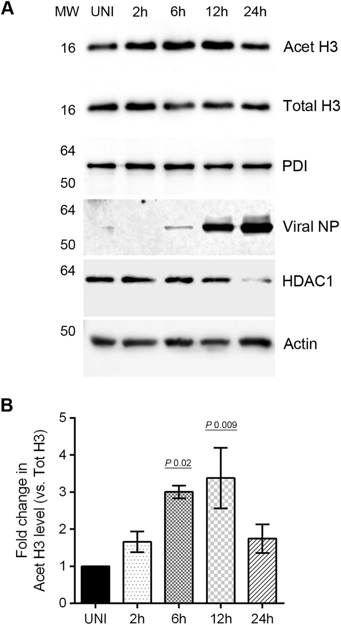
FIG 4.
IAV downregulates the activity of HDAC1. (A) MDCK cells (8 × 10⁵) were infected with PR8 at an MOI of 0.5 and harvested after 2, 6, 12, and 24 h of infection. Total cell lysates were prepared, and acetylated-histone H3 (Lys9) (Acet H3), total histone H3 (Total H3), PDI, HDAC1, actin, and NP were detected in uninfected (UNI) and infected (2, 6, 12, and 24 h) cell lysates by WB. Acet H3, an HDAC1 substrate, was detected as a marker of HDAC1 activity, and total H3, PDI, and actin were detected as loading controls. (B) Acet H3 and total H3 protein bands were quantified as Fig. 1C, and the amount of Acet H3 was normalized to the total H3. The normalized amount of Acet H3 in UNI sample was considered 1-fold for comparisons to 2-, 6-, 12-, and 24-h samples. The data presented are means ± the standard errors of the means of three independent experiments; the P value was calculated by using one-way ANOVA. MW, molecular weight.

