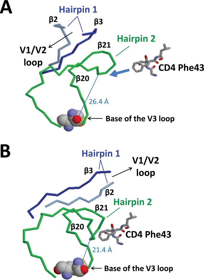
FIG 12.
Comparison of the gp120 bridging sheet in the CD4-unliganded and CD4-bound conformations. The structures of the bridging sheet elements in the crystal of CD4-unliganded trimer (PDB accession number 4NCO) (A) and the crystal of CD4-bound gp120 core (PDB accession number 1G9M) (B) were compared. Hairpin 1 (β2 [blue]-β3 [gray]), hairpin 2 (β20-β21 [green]), and the orientation of the V1/V2 loops are indicated (V1/V2 loops are not shown in the unliganded crystal and are truncated in the CD4-bound crystal). Residue Phe43 of CD4 is also shown (schematically in the unliganded crystal). The distance between carbon α of Trp422 in the tip of hairpin 2 and carbon α of Cys297 at the base of the V3 loop is also indicated (the cysteine residues at the base of the V3 loop are shown in space-fill for orientation). (A) In the unliganded crystal, β3 of hairpin 1 forms hydrogen bonds with β21 of hairpin 2, and the V1/V2 loops are oriented toward the trimer apex. (B) After binding to CD4, hairpin 1 twists and flips. Now, β2 of hairpin 1 forms hydrogen bonds with β21 of hairpin 2, and the V1/V2 loops are oriented away from the trimer apex. In addition, the distance between the tip of hairpin 2 and the base of the V3 loop is decreased by 5 Å.

