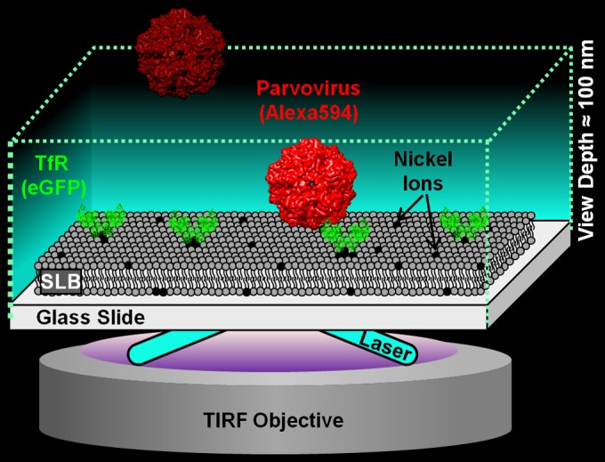
FIG 1.

Schematic of the single-particle tracking (SPT) binding assay. The device was set up on a 100×, 1.46-numerical-aperture, oil-immersion objective in a total internal reflection fluorescence (TIRF) microscope (Carl Zeiss; Model Axio Observer Z1). The TIRF microscope used here produces an evanescent wave that illuminates only a 100-nm-deep region away from the glass surface, which is where the virus-TfR binding interaction occurs, thus ignoring virus particles floating in the bulk solution. The eGFP-labeled TfRs are tethered to the supported lipid bilayer (SLB) via nickel-His interactions. The Alexa 594-labeled virus is detected with a 561-nm laser, whereas the eGFP-labeled TfR is detected using a 488-nm laser. The CPV structure is from VIPERdB based on PDB identifier 1P5W. The TfR structure is from PDB identifier 1CX8. Note that the CPV and TfR structures are used only for illustrative purposes and deviate from the virus capsid and receptor structures used in this work.
