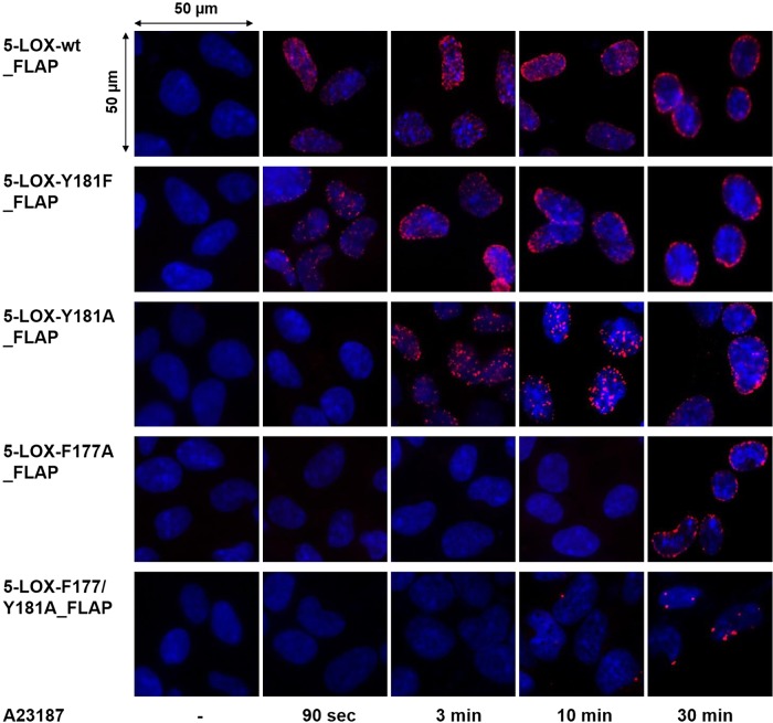
Figure 4.
Time-resolved in situ protein–protein interaction of 5-LOX-wt and mutants with FLAP. In situ PLA, using proximity probes against 5-LOX and FLAP, was performed in designated cell lines upon stimulation with A23187 (2.5 µM) for indicated time points. DAPI (blue) was used to stain nucleus, and in situ PLA signals (red dots) visualize 5-LOX/FLAP interaction. Scale bar, 50 µm. Results are representative of 100 individual cells analyzed in 3 independent experiments.

