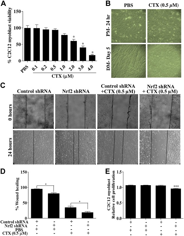
Figure 10.
CTX treatment hampers C2C12 myoblast differentiation and wound healing. A) Graph illustrates the percentage C2C12 cells viability measured by MTT assay in PBS and CTX treatment. B) Bright field light microscopy images showing the effect of 0.5 µM CTX treatment on C2C12 myoblast differentiation (×10 magnification). C) Artificial scratch wounds were created in Nrf2-silenced C2C12 monolayers, and the wound closure was captured by light microscope (×10 magnification). D) Graph represents the percentage of wound healing calculated by measuring the area of wound using ImageJ software. E) MTT cell viability assay of C2C12 myoblast transfected with control and Nrf2 shRNA for 48 h and treated with CTX (0.5 µM) for 24 h. Statistical significance was calculated by Student’s t test. *P < 0.05 between percent of wound healing of PBS and CTX-treated myoblast. ***P < 0.05 between control-shRNA+CTX vs. Nrf2-shRNA+CTX.

