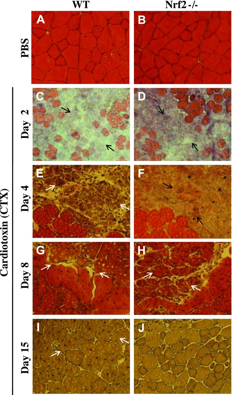
Figure 8.
Regeneration of CTX-induced TA muscle injury was delayed in Nrf2−/− mice. Bright field images of hematoxylin and eosin-stained cross sections of TA muscle injected with PBS (A, B) and post-CTX injury TA muscles collected on d 2 (C, D), d 4 (E, F), d 8 (G, H) and d 15 (I, J) of WT (A, C, E, G, I) and Nrf2−/− (B, D, F, H, J) mice (n = 4–5 mice/group). On d 2 postinjury, TA muscles of WT and Nrf2−/− mice show formation of edema and inflammatory infiltration (C, D) (black arrow). On d 4 (E, G) and d 8 postinjury (H), muscles show the presence of multinucleated myofibers (white arrow). On d 15 postinjury (I, J) larger cross-sectional and multinucleated myofibers can be seen in WT compared with Nrf2−/− mice (white arrow).

