Figure 9.
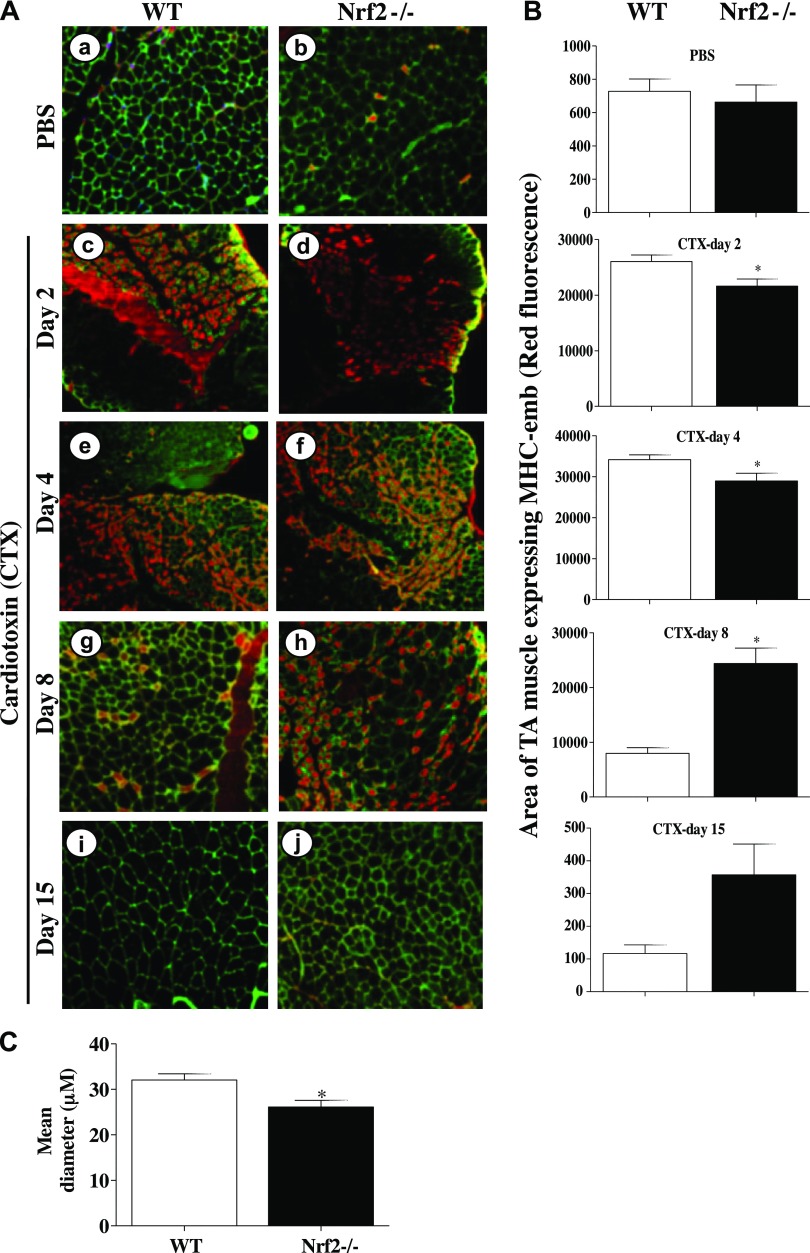
Delayed expression of MHC-emb in TA muscle of Nrf2−/− mice injured with CTX. A) Cross sections of regenerating TA muscle of WT (a, c, e, g, i) and Nrf2−/− (b, d, f, h, j) mice showing anti-MHC-emb (red fluorescence). Muscle cell plasma membrane and nucleus were counter stained with anti-laminin (green) and DAPI (blue), respectively. Cross section of PBS-injected TA muscles of WT and Nrf2−/− mice (a, b), and cross-sections at 2 d (c, d), 4 d (e, f), 8 d (g, h), and 15 d (i, j) following CTX injury of TA muscle of WT and Nrf2−/− mice. B) Average area of TA muscle expressing MHC-emb of n = 3 section of each group was determined using ImageJ software. C) Average diameter of ∼100 green positive myotubes per field (Ai, j) of n = 3 sections of each group was determined using ImageJ software. n = 3/d per group. Statistical significance was calculated by 1-way ANOVA. *P < 0.05 CTX-WT vs. Nrf2−/−.

