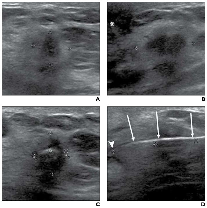
Fig. 1. 73-year-old woman with 14-mm invasive ductal carcinoma. Imaging sequence obtained during cryoablation procedure is shown.
A and B, Transverse (A) and sagittal (B) sonographic images were obtained at beginning of cryoablation procedure to quantify tumor size Calipers show maximal lesion diameters in both A and B; asterisk (B) denotes nipple.
C and D, After cryoprobe placement, transverse (C) and sagittal (D) sonographic images were acquired before initiation of freezing to confirm proper cryoprobe positioning. Carets show maximal radiuses from cryoprobe to mass periphery at 12:00, 3:00, 6:00, and 9:00 in C and distance from cryoprobe tip to distal lesion edge (left and center marks) is shown in D; arrows (D) denote cryoprobe, and arrowhead (D) identifies cryoprobe tip.

