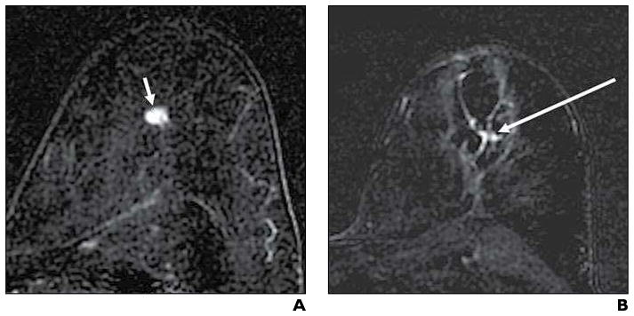
Fig. 4. 58-year-old woman with invasive ductal carcinoma with false-positive contrast-enhanced (CE) MRI results after cryoablation.
A and B, Initial enhanced axial subtraction CE-MR images before (A) and 31 days after (B) cryoablation show 9-mm enhancing malignant mass (arrow, A) and persistent focal enhancement (arrow, B) centrally within cryoablation area; although the latter finding was interpreted as suspicious for residual carcinoma, no residual malignancy was found at subsequent surgical resection.

