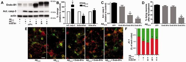
Figure 6.
Neuron-specific endophilin-B1 isoforms protect against amyloid-β (Aβ) neurotoxicity in vitro. (A) Postnatal cortical neurons were infected with lentivirus expressing either EGFP or endophilin-B1 isoforms for 3 days, then treated with 10 µM amyloid-β25-35 or reverse peptide amyloid-β35-25 for 24 h. Western blot analysis of endophilin-B1, activated caspase-3 (apoptosis marker), and actin (loading control) are shown. (B) Quantification of amyloid-β25-35-induced changes in the expression of endophilin-B1a, endophilin-B1b/c and activated caspase-3. Values are normalized to actin and expressed as per cent change relative to amyloid-β35-25 control. Amyloid-β25-35 induced an increase in activated caspase-3 and a decline in endophilin-B1b/c, but no change in endophilin-B1a (n = 4 separate experiments). *P < 0.05 versus amyloid-β35-25, Student’s t-test (n = 4). (C) Overexpression of neuron-specific endophilin-B1 isoforms endophilin-B1b and endophilin-B1c significantly decreased caspase-3 activation induced by amyloid-β25-35 (normalized to actin and relative to amyloid-β25-35/EGFP). *P < 0.05 versus amyloid-β25-35/EGFP, one-way ANOVA with Tukey post hoc test (n = 4). (D) Overexpression of endophilin-B1c, but not endophilin-B1b, reduced neuronal death induced by amyloid-β25-35. *P < 0.05 versus amyloid-β25-35/EGFP, one-way ANOVA with Tukey post hoc test (n = 3). (E) Cultured cortical neurons labelled with the JC-1 dye, an indicator of mitochondrial membrane potential, are shown. Healthy, polarized mitochondria emit red fluorescence, while damaged depolarized mitochondria show green fluorescence. Scale bar = 20 µm. (F) Quantification of JC-1 fluorescence intensity. *P < 0.05 versus amyloid-β35-25; #P < 0.05 versus amyloid-β25-35, one-way ANOVA with Tukey post hoc test (n = 4).

